. 2020 Jun 18;9:e59264. doi: 10.7554/eLife.59264

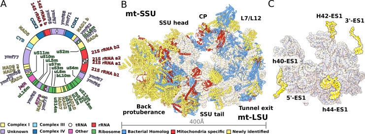
Figure 1. Structure of T. thermophila mitoribosome and newly identified proteins in the mitochondrial DNA.

(A) Schematic representation of the mitochondrial genome of T. thermophila with newly identified proteins labeled inside the circle. (B) The overall structure of the mitoribosome showing mitochondria specific and newly identified proteins. (C) Mitoribosomal rRNA showing expansion segments (relative to E. coli) in yellow.

Figure 1.

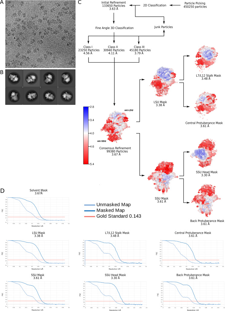
Figure 1—figure supplement 1. Electron microscopy data processing workflow.

Figure 1—figure supplement 1.

(A) Representative electron micrograph at 1.07 Å2/px. (B) 2D classes showing views of the T. thermophila mitoribosome. (C) Data processing steps with the final maps colored by local resolution. (D) Gold-standard Fourier shell correlation (FSC) curves for the final maps.
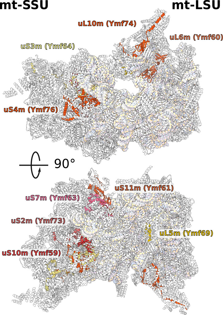
Figure 1—figure supplement 2. Positions of newly annotated proteins on the mitoribosome.

Figure 1—figure supplement 2.

Nine proteins with previously unknown function, which were named Ymf, have been identified as mitoribosomal proteins and modeled as shown.
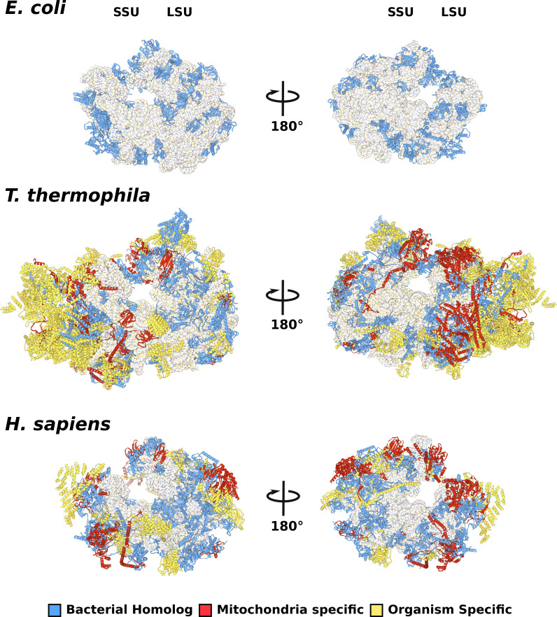
Figure 1—figure supplement 3. Comparison of evolutionary conservation for mitoribosomal proteins.

Figure 1—figure supplement 3.

The overall structures of E. coli (PDBID: 5MDZ), T. thermophila (PDBID: 6Z1P), and H. sapiens (PDBID: 3j9m) colored by protein conservation.
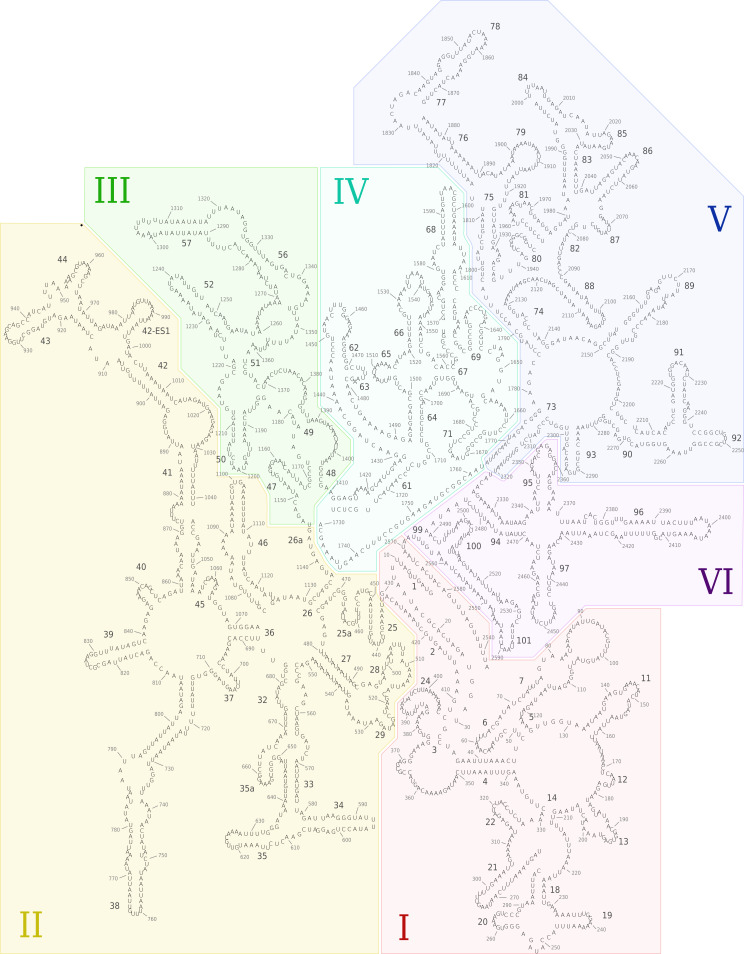
Figure 1—figure supplement 4. Secondary structure diagram of the T. thermophila LSU rRNA.

Figure 1—figure supplement 4.

The rRNA is colored by domain, and expansion segments are indicated. Fragmentation is found in H18.
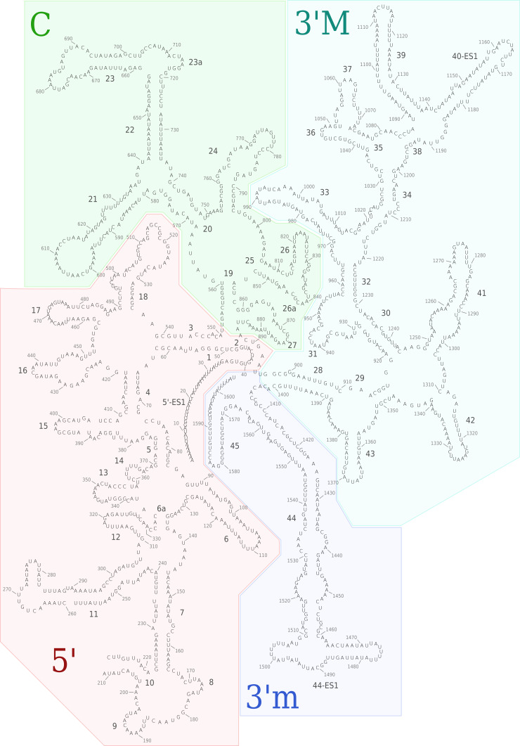
Figure 1—figure supplement 5. Secondary structure diagram of the T. thermophila SSU rRNA.

Figure 1—figure supplement 5.

The rRNA is colored by domain, and expansion segments are indicated. Fragmentation is found in h10.