Figure 6.

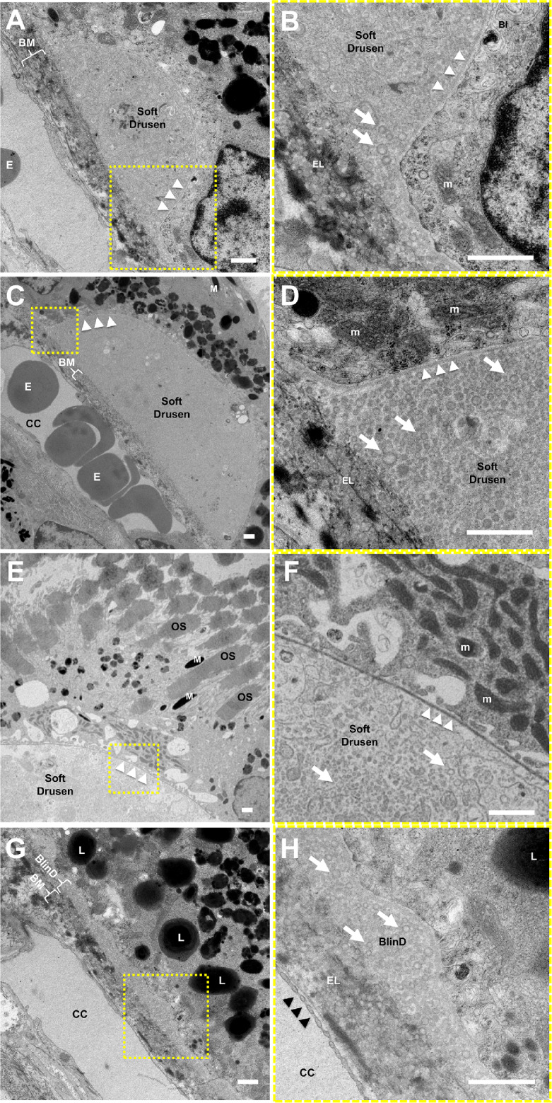
Ultrastructure of soft drusen in rhesus macaques. TEM images of (A, B) a small soft druse, (C–F) larger drusen, and (G, H) thin basal linear deposits (BlinD) located between the RPE basal lamina (white arrowheads) and Bruch's membrane (BM). The magnified views in B, D, F, and H correspond to the dashed-yellow areas in A, C, E, and G. The neurosensory retina is located at the upper right and the choroid/sclera are toward the lower left. The soft drusen in A–F and BlinD in G and H are both largely composed of vesicular particles with an electron-dense exterior and electron-lucent interior (arrows) consistent with partially extracted lipoprotein particles. Melanosomes (M), lipofuscin granules (L), and mitochondria (m) in RPE cells; overlying photoreceptor outer segments (OS); and erythrocytes (E) in the choriocapillaris (CC) can also be seen. The elastic layer (EL) of BM, scant RPE basal infoldings (BI), and fenestrations of the CC (black arrowheads) are better seen on the magnified views. Scale bars: 1 μm.
