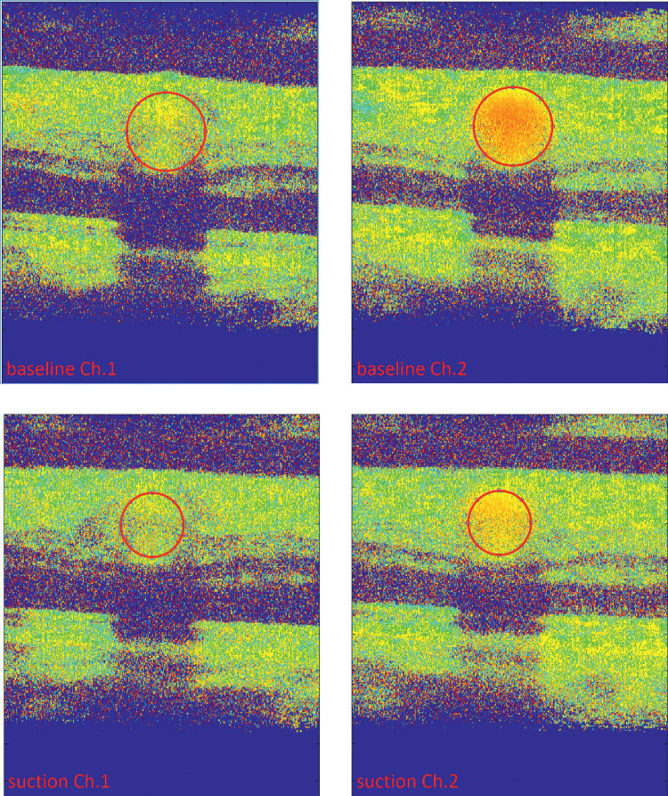
Figure 6.
Representative summed phase images of both channels as acquired with the bidirectional Doppler-OCT at baseline (top) and during the highest suction level (bottom). As for channel 1, the angle of the measurement beam was almost perpendicular to the vessel and the observed phase shift in channel 1 is only small. Vessel diameter was determined based on the channel with the higher phase shift (channel 2). The vessels of interest are marked in red and decrease in size upon suction.

