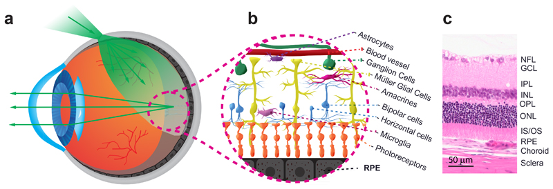
Fig. 1. Illumination of the retinal layers provided via transscleral illumination.
(a) Light transmission into the eye. The light is first transmitted through the sclera, the retina pigment epithelium (RPE), and the neuroretina. After travelling through the vitreous humor and the retina, it impinges on the RPE layer. The light is multiply scattered by the RPE. A part travels back and propagates through the neuroretina and its translucent cells, the vitreous, the eye lens, and, then, the anterior segment of the eye. (b) Artistic illustration of the translucent vascular, neuronal and glial cells of the retina. (c) Histological cut of the retinal layers, the choroid, and the sclera. GCL: ganglion cells layer; INL: inner nuclear layer; IPL: inner plexiform layer; IS/OS: inner and outer segments of the photoreceptors; NFL: nerve fiber layer; ONL: outer nuclear layer; OPL: outer plexiform layer.

