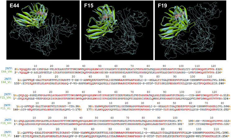
FIGURE 1.
The 3D complex of RS2-1G9 F(ab′)2 fragment and N-Acyl-L-homoserine lactone analog (PDB:2NTF) was superimposed by the HuscFv-E44 (left), HuscFv-F15 (middle), and HuscFv-F19 (right) using CLICK: http://mspc.bii.a-star.edu.sg/click (Upper panel). One antigen-binding site of the mAb RS2-1G9 F(ab′)2 fragment (VH and VL domains, shown in blue) was superimposed by the HuscFvs (green). The trace illustration is the remaining portion of the RS2-1G9 F(ab′)2. Lower panel, the superimposed amino acids of the RS2-1G9 antigen-binding site (2NTF) and the VH and VL of HuscFv-E44, HuscFv-F15, and HuscFv-F19, are shown in red alphabets.

