FIGURE 3.
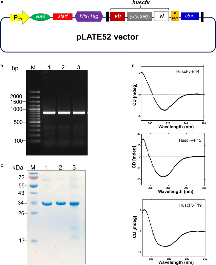
Production and characterization of HuscFvs to 3O-C12-HSL. (A) Schematic diagram of the inserted DNA construct in pLATE52 where the DNA sequence coding for HuscFv (vh-linker-vl) was flanked with DNA sequences of 6 × His at the 5′ end and E-tag at the 3′ end. (B) Amplicons of huscfv-LIC fragments (∼ 850 bp) for sub-cloning into pLATE52 vector. M, 100 bp-plus DNA ladder; 1–3, huscfv-LIC amplicons of three representatives transformed NiCo21(DE3) E. coli clones. Numbers at the left are DNA sizes in bp. (C) Stained SDS-PAGE-separated purified recombinant HuscFvs. M, protein standard; 1–3, purified HuscFv-E44, HuscFv-F15 and HuscFv-F19, respectively. Numbers at the left are protein masses in kDa. (D) CD spectra of the refolded HuscFv-E44, HuscFv-F15, and HuscFv-F19.

