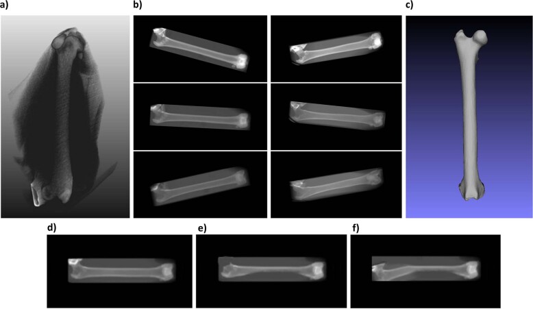
Fig. 1. Dataset.
a 3D CT DICOM file. b Examples of X-ray images artificially generated from 3D CT DICOM data. Images in the left column of b were generated from the same bone. Images in the right column of b were generated from a second bone. c Bone mesh (surface model) extracted from the 3D CT DICOM file. d Artificial X-ray image of a healthy bone. e Artificial X-ray image of a deformed bone. f Artificial X-ray image of a strongly deformed bone.

