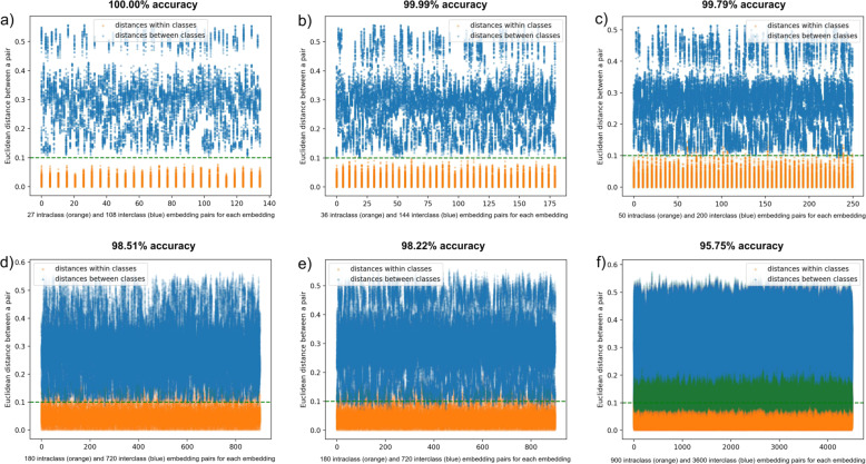
Fig. 4. Distribution of pairwise Euclidean distances between embeddings of 4500 artificial X-ray images generated from five bones.
The Triplet network was not trained on any images of these bones. a Pairwise embedding distances between images depicting bones from an angle which did not deviate from the standard anteroposterior or mediolateral (AP/ML) view by more than 4° in any direction. Radiation energy interval: 146–158 keV. b Maximum deviation from AP/ML view: 4°. Radiation energy interval: 140–158 keV. c Maximum deviation from AP/ML view: 7°. Radiation energy interval: 140–158 keV. d Maximum deviation from AP/ML view: 22° around the bone′s longitudinal axis and 4° around an axis perpendicular to image plane. Radiation energy interval: 140–158 keV. e Maximum deviation from AP/ML view: 4° around the bone′s longitudinal axis and 22° around an axis perpendicular to image plane. Radiation energy interval: 140–158 keV. f Maximum deviation from AP/ML view: 22°. Radiation energy interval: 140–158 keV. Overlapping dots are marked green.

