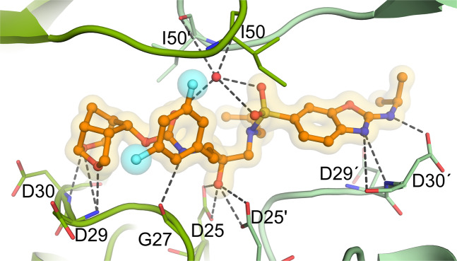
Figure 4.
The X-ray crystal structure of GRL-063 bound to the active site of wild-type HIV-1 protease (PRWT). Carbon atoms of GRL-063 are shown in orange and subunits of PRWT in green tones. Nitrogen, oxygen, sulfur, and fluorine atoms are shown in blue, red, yellow, and cyan, respectively. Hydrogen bond interactions that took place between GRL-063 and the protease residues are indicated by gray dashed lines, and two fluorine atoms are highlighted by cyan spheres.

