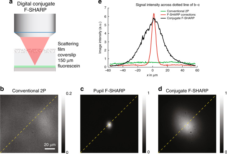
Fig. 2. Conjugate F-SHARP of a uniform fluorescein sample through a thin scattering layer.
Imaging comparison of a uniform fluorescein layer placed 150 μm away from a thin (≲50 µm) scattering layer: (a) conventional 2P microscopy (b), pupil F-SHARP imaging (c) and conjugate F-SHARP (d). Intensity profile of the fluorescence image captured with the three configurations along the dotted line of b–d (e). Images in b through d were acquired with the same laser power and pixel dwell time and normalized to the maximum value of the 3.

