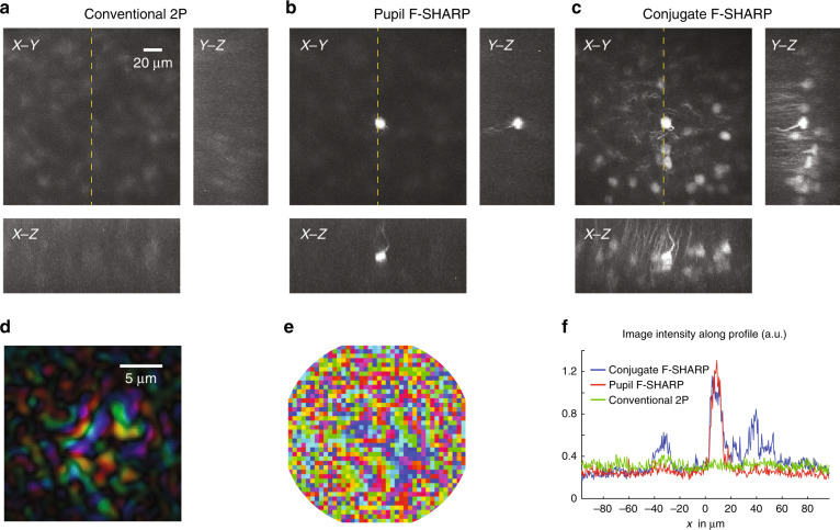
Fig. 4. Imaging through the thinned skull of a densely labelled Rbp4-Cre/Ai9 mouse 360 μm below the brain surface.
Maximum intensity projection of a 190 × 190 × 80 μm3 volume using conventional 2P (a), pupil F-SHARP (b) and conjugate F-SHARP (c). As in Fig. 3, the images in a–c are presented on the same colour scale, and saturation corresponds to 0.5 of the maximum signal. The measured E-field PSF at the centre of the image (d) and the corresponding correction pattern assigned to the wavefront shaper (e) demonstrate a scattered PSF with a smaller number of modes compared to Fig. 3. The high variability in the skull thinning process can lead to these differences. The conjugate F-SHARP module allows us to increase both the signal intensity and the resolution over an extended FOV compared with conventional 2P and F-SHARP (f).

