Fig. 5.
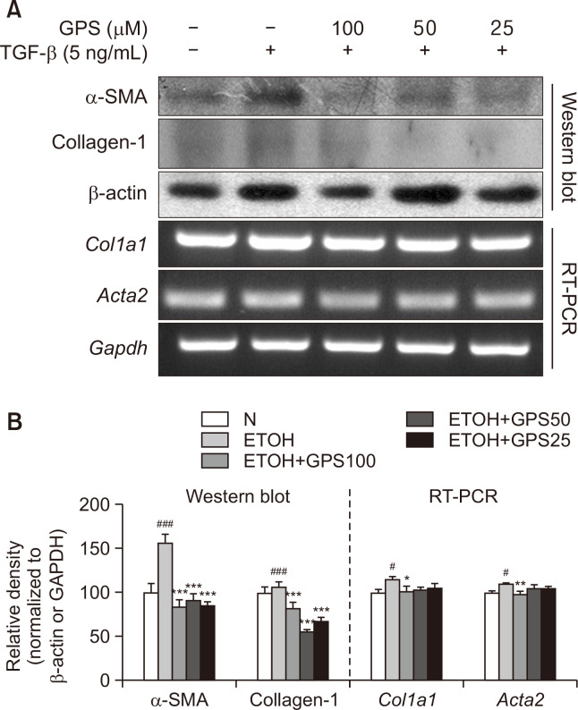
GPS suppressed TGF-β-stimulated HSC activation. LX-2 cells were pretreated with GPS for 1h in the presence of TGF-β (5 ng/mL) for 24 h. (A) Protein expression of α-SMA and collagen I was analyzed by western blotting. mRNA expression of Acta2 (encoding α-SMA) or Col1a1 (encoding collagen I) was analyzed by RT-PCR. (B) Densitometric tracing analysis was done for each western blot band or RT-PCR band and normalized to β-actin or GAPDH. #p<0.05, ###p<0.001, significantly different from untreated cells; *p<0.05, **p<0.01, ***p<0.001 significantly different from TGF-β-stimulated cells. All histograms represent the mean ± SD of at least three independent assays.

