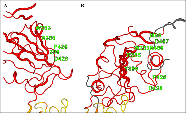
Fig. 4.
Images showing the differences between paired C-alpha residues from chain A and chain B of the spike protein coloured according to their extent of difference using 2StrucCompare [10]. Residues coloured in red indicate differences in paired positions greater than 5 Å from each other between the two chains. a. Shows the shape and position of chain A in the “up” position. b. Shows the same orientation for chain B in the “down” position. Some residues are labelled to indicate approximately where the pocket residues are in the two positions of these chains, and in B three of these residues lie on the strand coloured dark grey indicating that this is not seen in chain A

