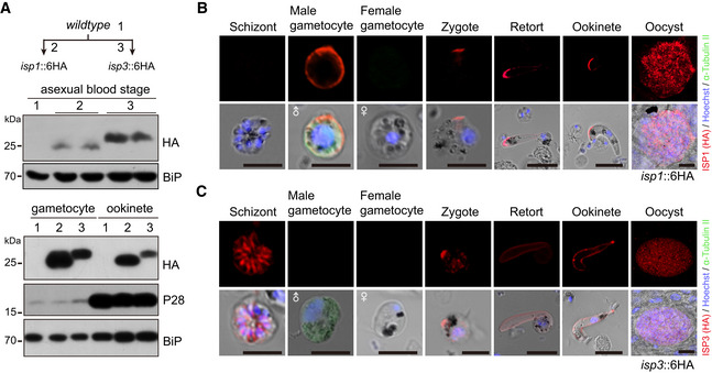
Figure EV2. Stage expression and cellular localization of ISP1 and ISP3.

-
AWestern blot of ISP1 and ISP3 in asexual mouse blood stages, gametocytes, and ookinetes of the isp1::6HA and isp3::6HA parasites. ER protein BiP as loading control. Two lanes in blot are replicates from the same sample.
-
B, CIFA of ISP1 (B) and ISP3 (C) in mouse and mosquito stages of the isp1::6HA and isp3::6HA parasites, respectively. Purified gametocytes were stained with antibodies against HA and α‐tubulin II (male gametocyte‐specific). Scale bar = 5 μm.
Source data are available online for this figure.
