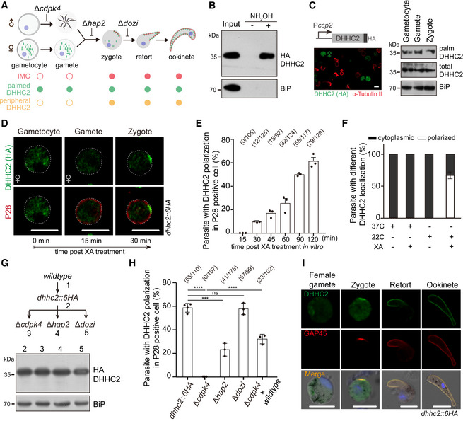
Figure 6. Newly assembled IMC is a polarity patch for DHHC2 targeting in fertilized zygote.

- Schematic of IMC assembly during gametocyte to ookinete. IMC assembly initiates in apical and extends along protrusion of the fertilized zygotes.
- Endogenous DHHC2 is palmitoylated in the dhhc2::6HA gametocytes.
- Episomally expressed DHHC2 driven by promoter of ccp2 gene is palmitoylated in female gametocytes, female gametes, and zygotes of WT parasites. Left panel shows the expression of HA‐tagged DHHC2 in female gametocyte by co‐staining with antibodies of HA and α‐tubulin II (male gametocyte‐specific). Scale bar = 5 μm.
- IFA of DHHC2 and P28 in female gametocytes, female gametes, and zygotes of the dhhc2::6HA parasite. Gametocytes were stimulated with XA and 22°C in vitro. P28 is expressed specifically in female gametes and zygotes. Scale bar = 5 μm.
- Quantification of parasites with DHHC2 polarization in P28‐positive cells during gametocyte‐to‐zygote differentiation in (D). x/y: the number of cells with DHHC2 polarization and the number of cells analyzed. Values are means ± SD from three independent tests.
- No polarization of DHHC2 in the dhhc2::6HA gametocytes with only either XA or 22°C stimulation. Three replicates are performed, and values are means ± SD.
- No change in DHHC2 expression in zygotes of the dhhc2::6HA parasite‐derived mutants with individual gene disruption of cdpk4, hap2, and dozi.
- Quantification of parasite with DHHC2 polarization in P28‐positive cells. Parasites are indicated in (G) or from a genetic cross (dhhc2::6HA/cdpk4 and WT). x/y: the number of cells with DHHC2 polarization and the number of cells analyzed. Values are means ± SD from three tests, two‐tailed t‐test, ***P < 0.001, ****P < 0.0001, ns, not significant. Cell images in Appendix Fig S4.
- Expression and localization of DHHC2 and GAP45 during female gamete‐to‐ookinete development of dhhc2::6HA parasite. GAP45 is an IMC marker and is only detected in zygote after fertilization, but not gametes. Scale bar = 5 μm.
Source data are available online for this figure.
