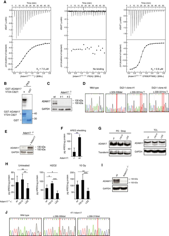
Figure EV4. Regulation of ADAM17 by PP2A‐B56.

- ITC measurements of the binding of B56α to the indicated ADAM17 peptides.
- In vitro phosphorylation of GST and GST‐ADAM17 with PKA.
- Western blot of ADAM17 expression in the parental wild‐type (wt) DLD‐1 cell line and ADAM17 knockout (ADAM17−/−) clones #1 and #2.
- Sanger sequences of the edited coding region in ADAM17 of DLD‐1 wt and ADAM17−/− clones #1 and #2.
- Re‐expression of ADAM17 in ADAM17−/− (clone #1) cells, determined by Western blot.
- Amphiregulin (AREG) shedding measured by ELISA of conditioned media from ADAM17−/− cells (clone #1) with or without ADAM17 re‐expression (A17−/−+A17). Two‐sided, unpaired Student's t‐test was applied to test for significant differences **P < 0.01. Mean and standard deviation indicated from three independent experiments.
- Western blot of ADAM17 in streptavidin pull‐downs (PD: Strep) and total cell lysates (TCL) from cell surface biotinylated DLD‐1 ADAM17−/− cells (clone #1) re‐expressing ADAM17 variants (wt, I761 or LEE).
- Amphiregulin (AREG) shedding measured by ELISA in conditioned media from untreated, H2O2 treated, or radiated DLD‐1 ADAM17−/− cells (clone #2) re‐expressing ADAM17 variants (wt, I761 or LEE). Two‐sided, unpaired Student's t‐test was applied to test for significant differences *P < 0.05, **P < 0.01 and ***P < 0.001. Mean and standard deviation indicated from at least three independent experiments.
- Western blot of ADAM17 expression in the parental 4T1 cells line (wt) and 4T1 ADAM17 knockout clone (A17−/−).
- Sanger sequences of the edited coding region in Adam17 of the 4T1 wt and A17−/− cell lines. GAPDH was used as an internal loading control in all Western blots. Shedding data represent average values ± SEM of at least 3 independent experiments.
Source data are available online for this figure.
