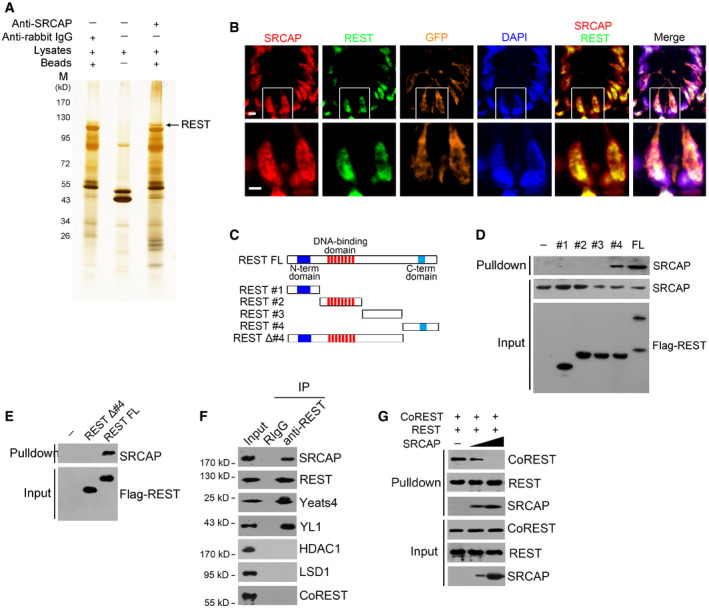
Nuclear extracts of murine intestinal crypts were immunoprecipitated with anti‐SRCAP antibody or rabbit IgG followed by ADASDS–PAGE, sliver staining, and mass spectrometry.
SRCAP and REST were stained in murine intestinal crypts. Scale bar, 10 μm.
A schematic diagram of full length and truncated fragments of REST protein was shown.
Flag‐tagged REST fragments were expressed in 293T and purified by immunoprecipitation with anti‐Flag antibody. REST proteins were incubated with crypt lysates for pulldown followed by immunoblotting.
C‐terminal domain of REST was sufficient and essential for SRCAP association by pulldown assay.
Intestinal crypt lysates were incubated with anti‐REST antibody, followed by immunoblotting with indicated antibodies.
Intestinal crypt lysates were incubated with anti‐REST antibody and protein A/G beads in 4°C overnight. Recombinant SRCAP protein was added with different concentrations, followed by immunoblotting.
Data information: All data are representative of four independent experiments.

