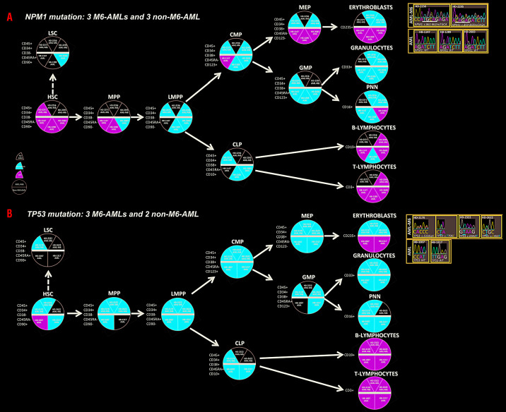
Figure 1.
Distribution of NPM1 and TP53 mutations in hematopoietic compartments of erythroid acute myeloid leukemia (M6-AML) and non-M6-AML. (A) Three M6-AML and three non-M6-AML sequenced for NPM1. Each circle represents a cell at a different level of the classical hematopoietic hierarchy from the most immature to mature population. The immunophenotype of these cells corresponds to the cell sorting performed. Each cell/circle is separated in two by a bold black line with M6-AML above and non-M6-AML below. Inside each cell/circle, each slice corresponds to a Sanger-sequenced case with NPM1 mutation: red means mutated, green non-mutated and white no data. Results of NPM1 sequencing in erythroblasts are indicated to the right. M6-ALM: erythroid acute myeloid leukemia; AML: acute myeloid leukemia; HSC: hematopoietic stem cell, LSC: leukemic stem cell; MPP: multipotent progenitor; LMPP: lymphoid-myeloid pluripotent progenitor; CMP: common myeloid progenitor; CLP: common lymphoid progenitor; MEP: megakaryocyte erythroid progenitor; GMP: granulocyte-monocyte progenitor. (B) Three M6-AML and two non-M6-AML sequenced for TP53. Symbols as in (A).

