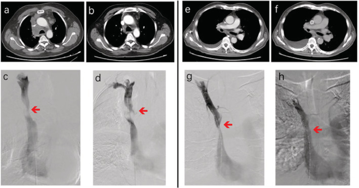
Figure 1.

The pre‐ and postoperative images of the two patients with superior vena cava syndrome (SVCS). (a–d) Patient 1 and (e–f) patient 2. (a and e) Preoperative computed tomography (CT) scan images of both patients; and (b and f) postoperative CT scan images of both patients. (c and g) Preoperative angiography of both cases; and (g and h) postoperative angiography of both cases.
