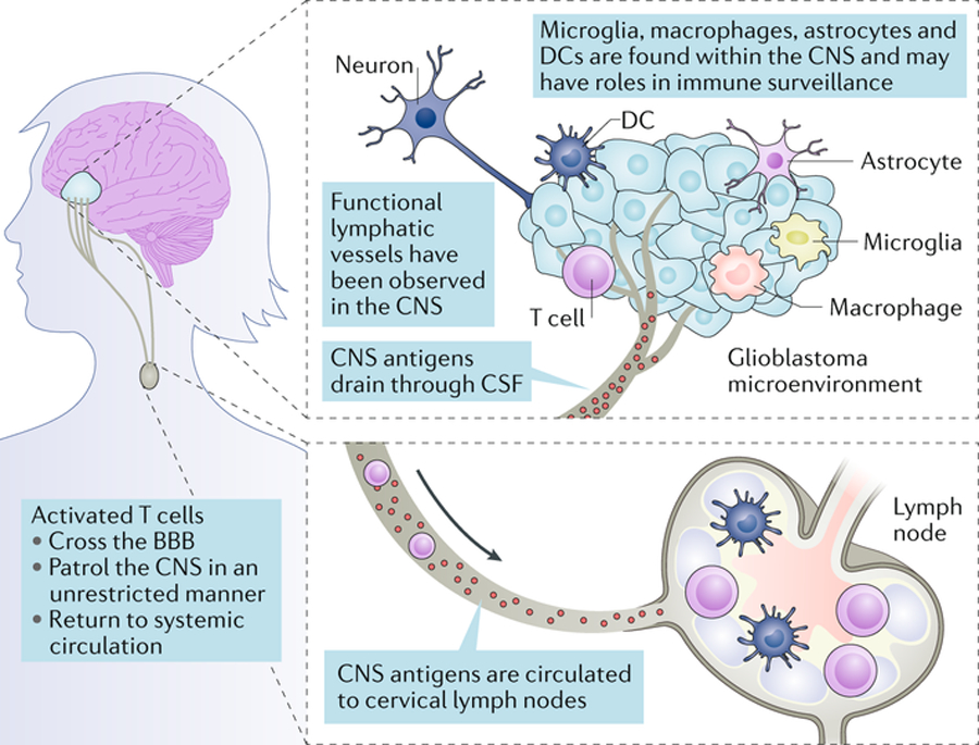
Fig. 1. Immune privilege in the brain.

Historically, the central nervous system (CNS) was considered to be isolated immunologically. Features that contributed to this understanding were the presence of tight junctions in the blood–brain barrier, the absence of a classic lymphatic drainage system, and empirical data showing the ability of the CNS to target foreign tissues with minimal inflammatory responses. However, today the concept of immune privilege has been partially redefined. It is now clear that there are functional lymphatic vessels in the CNS, and that antigen-presenting cells (APCs) of varied types exist within the CNS, including microglia, macrophages, astrocytes and classic APCs such as dendritic cells (DCs). It is now known that the CNS is not isolated from activated T cells, which can patrol these compartments in an unrestricted manner, and that CNS antigens can be presented locally or in the draining cervical lymph nodes. While the immune system in the CNS remains different, it is not incapable. CSF, cerebrospinal fluid.
