Fig. (3).

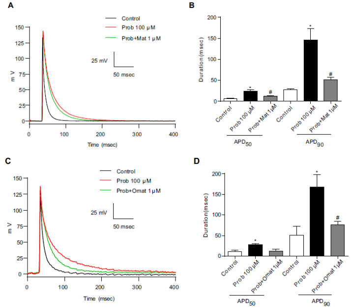
The effects of matrine and oxymatrine on the prolongation of APD by probucol in neonatal cardiac myocytes. (A) and (C) Representative action potential traces from control (black line), probucol (red line) and probucol/1 μM matrine or 1 μM oxymatrine groups (green line) in neonatal cardiac myocytes. (B) and (D) The APD50 and APD90 were prolonged by probucol and recovered by 1 μM matrine or 1 μM oxymatrine. n=5, * p <0.05 vs the control and # p <0.05 vs probucol. (A higher resolution / colour version of this figure is available in the electronic copy of the article).
