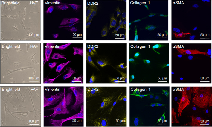
Fig. 1.

Morphological and immunocytochemical fibroblast identification. Representative brightfield and immunofluorescence images of the fibroblast markers vimentin, DDR2, collagen 1, and αSMA. The nuclei were stained with DAPI (blue). Upper panel) HVFs. Mid panel) HAFs. Lower panel) PAFs. The scale bars equal 50 µm.
