
FIG. 4.
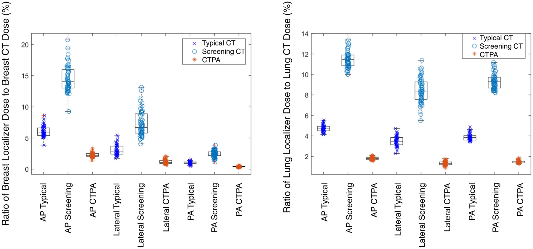
Breast dose (left) and lung dose (right) for a population of patients (n = 57) from the three localizer protocols (AP, lateral, and PA) used in this study expressed as a percentage of breast dose and lung dose for both a typical CT protocol dose (x), a low-dose lung cancer screening protocol (o), and a high-dose CTPA protocol (*). Outlier data points are shown with a red “+” symbol. The typical CT protocol dose scan had CTDIvol = 4.1 mGy, low-dose lung cancer screening scan had CTDIvol = 1.7 mGy, and the CTPA protocol had CTDIvol = 10.8 mGy.