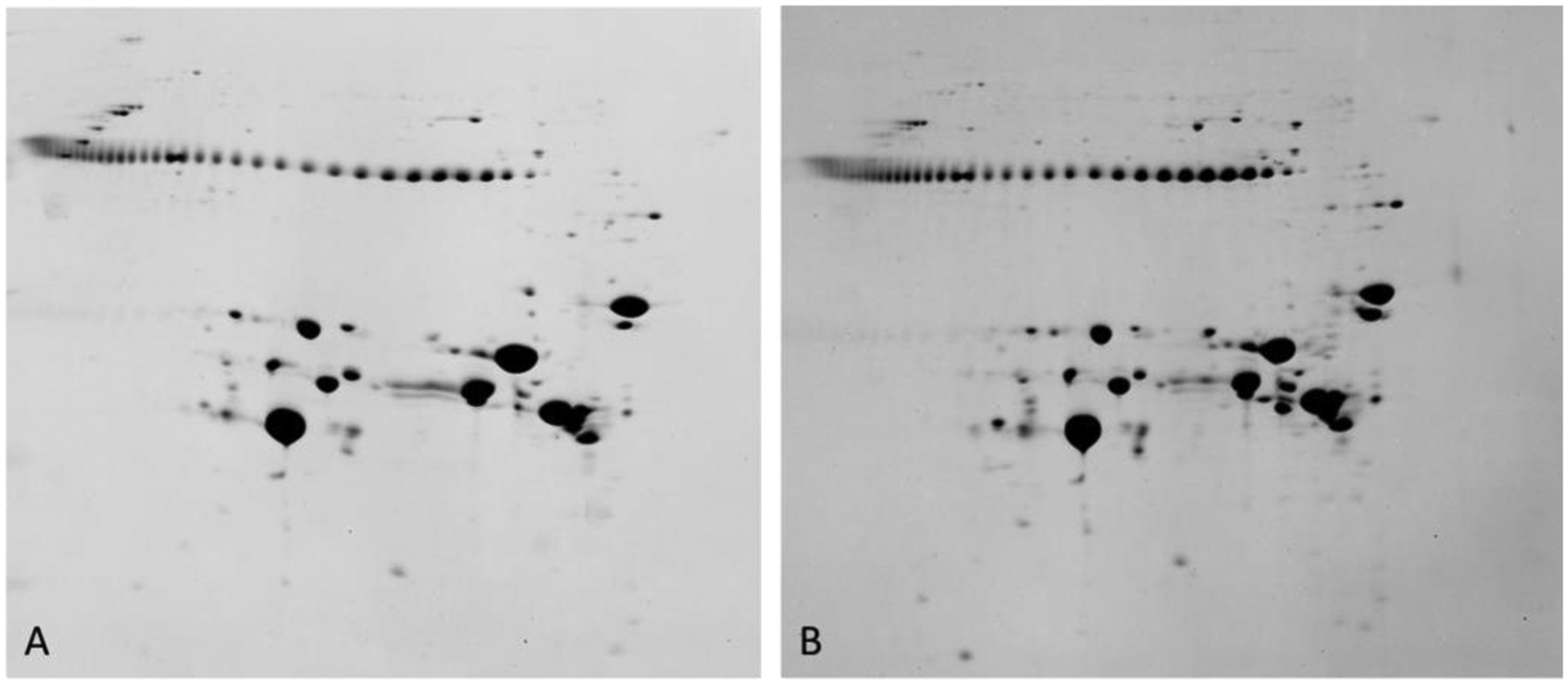
Figure 16. 2DE analysis of a 22d human cataractous lens.

2DE patterns of human lens proteins observed from the clear region (left) and cortical cataract region (right) of a 22 day old human lens. These data are for total protein including water soluble and water insoluble proteins. The string of protein spots at Mr 42k are pI markers. Proteins on the gel range from about pI 3 (left) to about pI 8.9 (right). Samples were solubilized in 9M urea, 2% NP40, 10mM DTT, 2% ampholytes (Resolyte pH 3.5–10). (Garland, unpublished data).
