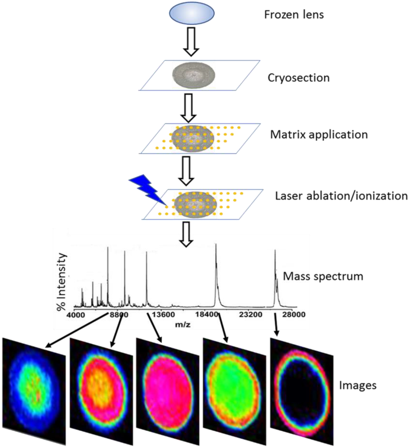
Figure 3. MALDI imaging mass spectrometry workflow.

Schematic diagram of workflow for imaging MALDI imaging mass spectrometry of frozen lens tissue showing major steps in the procedure including: cryosectioning, matrix application, laser ablation/ionization, data acquisition, and image generation.
