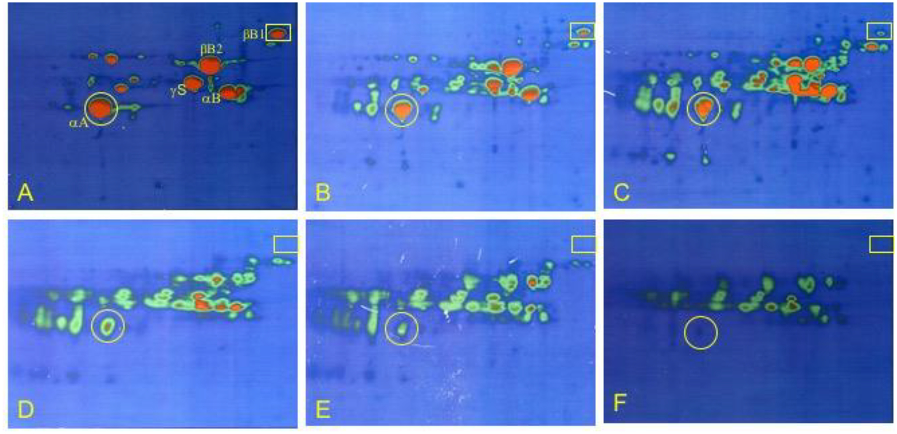
Figure 5. 2DE analysis of human lens regions.

Psuedo colored images of 2DE separation of 5 cortical shells of fiber cells and the nuclear region of a 45y lens. Left to right: outer cortical layers (panels A-E) with successive cortical fibers inward until the lower right image is of the lens nucleus (panel F). Protein concentration is from high (red) to low (green). The original locations of αA- and βB1-crystallin are indicated by yellow circles and rectangles, respectively. Samples were solubilized in 9M urea, 2% NP40, 10mM DTT, 2% ampholytes (Resolyte pH 3.5–10). Proteins were separated based on isoelectric point using 18 cm non-linear, immobilized pH 3–10 gradients for 1st dimension isoelectric focusing (separation from acidic on the left to basic on the right) and based on size using SDS-PAGE in the second, vertical, dimension (18 × 25 cm gels). Each image represents total protein, water soluble and water insoluble, for all samples including the lens nucleus. Crystallin species migrated between pI 8.6 and 5.
