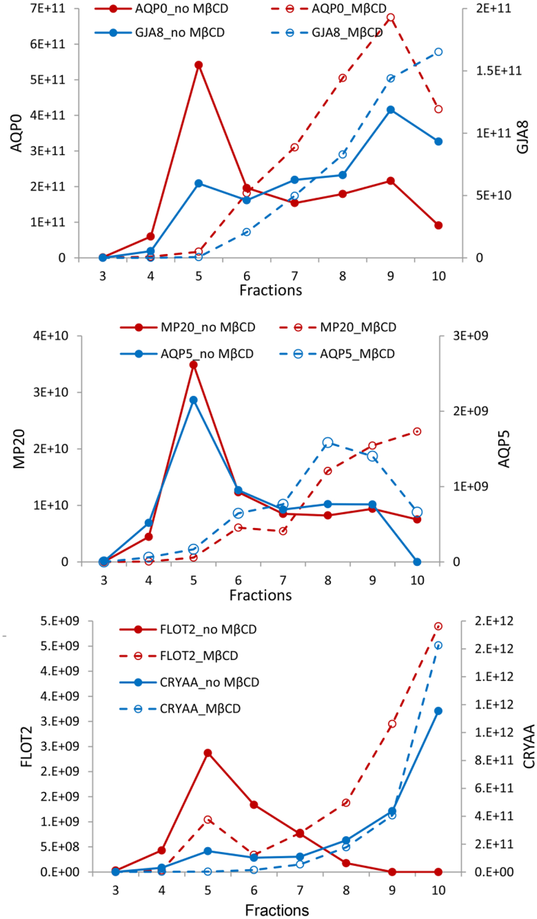
Figure 6. Identification of bovine lens lipid raft proteins.

Fractionation of lens membrane proteins in a sucrose density gradient indicating lipid raft domains (fractions 3–6) containing AQP0 and connexin 50 (GJA8) (top), MP20 and AQP5 (middle) and Flotillin-2 and αA-crystallin (bottom). The addition of methyl-β-cyclodextrin reduces lipid raft domains and shifts contents to later eluting fractions (dashed lines). The y-axes represent peak area intensities for corresponding peptides from each protein in arbitrary units. Figure from (Wang and Schey, 2015).
