Fig. 1.
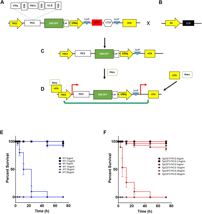
Design of the CETI-PIC3 line. (A) The genetic construct of the CETI-PIC3 line. A tetracycline-on (Tet-on) system is used to induce the three cytokines of interest. An H2B-GFP cassette is placed downstream of the cytokines as a marker to visually show that the cytokines have been induced. (B) This model takes advantage of Cre-lox systems to induce cytokines in a tissue-specific manner. (C) Any tissue-specific promoter driving a Cre cassette can be used to induce tissue-specific inflammation in this model after excision of the stop codon downstream of the RFP cassette. (D) When CET-PIC3 fish are crossed to a tissue-specific Cre and subsequently treated with doxycycline, there is dose-dependent induction of the cytokines in a tissue-specific manner. (E) Survival curve for wild-type (WT) embryos treated with different doses of doxycycline (0-50 µg/ml) for different time periods (0-72 h). (F) Survival curve for CETI-PIC3 embryos treated with different doses of doxycycline (0-50 µg/ml) for different time periods (0-72 h). There is no difference in survival for WT versus CETI-PIC3 doxycycline-treated embryos. n=10 embryos per dose/time. In all figures, data are mean±s.e.m.

