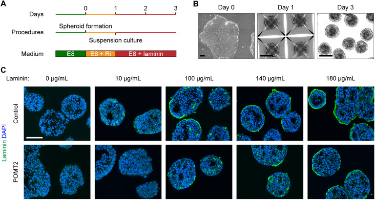
Fig. 6.
Accumulation of exogenous laminin on endoderm-free embryoid bodies. (A,B) Schematic and phase-contrast images of endoderm-free embryoid body culture protocol. Feeder-free hiPSCs were seeded on day 0 in Microwell plates to form spheroids of roughly 1000 cells by day 1. The spheroids were transferred to suspension culture supplemented with laminin for 48 h. (C) Immunohistochemistry demonstrating the effect of increasing laminin concentration on endoderm-free embryoid bodies. Embryoid bodies were supplemented with varying concentrations of laminin on day 1 and collected for analysis 48 h later. Two independent cell culture replicates were performed per cell line. Scale bars: 100 µm.

