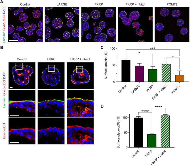
Fig. 7.
Patient-specific differences in laminin accumulation and response to ribitol treatment. (A) Immunohistochemistry on day 3 endoderm-free embryoid bodies supplemented for 48 h with exogenous laminin. Scale bar: 200 µm. (B) High-magnification images to assess the effect of ribitol treatment on the localization and glycosylation of αDG in FKRP patient embryoid bodies. Scale bar: 25 µm. (C) Quantification of the percentage embryoid body surface area covered by laminin: control, 66.6±4.7%; LARGE, 48.4±7.3%; FKRP, 38.2±8.3%; FKRP+ribitol, 53.8±6.3%; POMT2, 20.5±5.2%. Values expressed as mean±s.e.m. Three controls and two clones per patient were analyzed across n=12 (control), n=6 (LARGE and POMT2), and n=9 (FKRP and FKRP+ribitol) independent cell culture differentiations. Post hoc analysis with one-way ANOVA and Tukey's correction for multiple comparisons: *P<0.05, ***P<0.001. (D) Quantification of glyco-αDG staining intensity at the embryoid body surface: control, 100.0±4.2%; FKRP, 44.5±3.5%; FKRP+ribitol, 107.2±3.4%. Values normalized to a percentage of the controls and expressed as mean±s.e.m. Two controls and two FKRP clones were analyzed across three cell culture differentiations to measure n=54 surface regions of 50 µm length per condition. Post hoc analysis with one-way ANOVA and Tukey's correction for multiple comparisons: ****P<0.0001.

