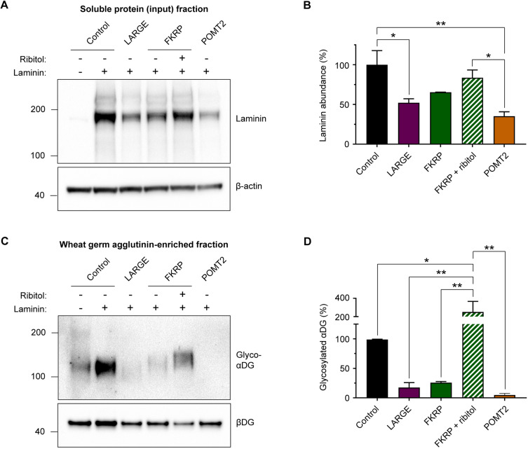
Fig. 8.
Western blot analysis of endoderm-free embryoid bodies. (A) Representative western blot measurement of laminin abundance in the input soluble protein fraction (not wheat germ agglutinin-enriched) from culture lysates of day 3 endoderm-free embryoid bodies. (B) Quantification of laminin band intensity: control, 100.0±17.8%; LARGE, 52.2±4.9%; FKRP, 65.4±0.2%; FKRP+ribitol, 83.6±5.7%; POMT2, 35.2±3.2%. Values expressed as mean±s.e.m. of n=3 independent cell culture replicates. Each sample was normalized to β-actin, and all samples are plotted as a percentage of control. Post hoc analysis with one-way ANOVA and Tukey's correction for multiple comparisons: *P<0.05, **P <0.01. (C) Representative western blot of glyco-αDG in wheat germ agglutinin-enriched culture lysates from day 3 endoderm-free embryoid bodies. (D) Quantification of glyco-αDG band intensity: control, 100.0±0.6%; LARGE, 17.6±8.3%; FKRP, 25.9%±1.9%; FKRP+ribitol, 250.8±67.3%; POMT2, 4.9±1.5%. Values plotted as mean±s.e.m. of n=3 independent cell culture replicates. Each sample was normalized to βDG, and all samples are expressed as a percentage of control. Post hoc analysis with one-way ANOVA and Tukey's correction for multiple comparisons: *P<0.05, **P<0.01.

