Fig. 1.
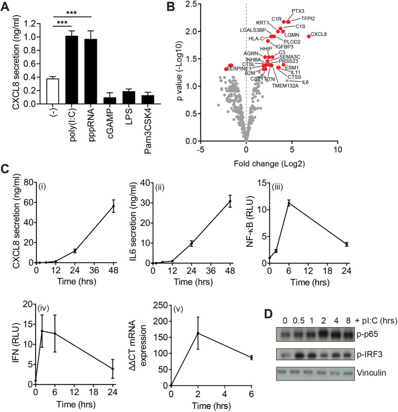
RPE cells show a robust TLR3 and RIG-I response. (A) CXCL8 secretion from RPE cells stimulated with the indicated ligands. Bars depict mean±s.e.m. of ≥n=4 independent experiments. ***P<0.001 (one-way ANOVA and a Bonferroni post-hoc test). (B) Volcano plot of fold change versus adjusted P-value from SILAC secretome experiments (n=3). Red points show a significant P-value change (P<0.05) in the poly(I:C)-stimulated versus unstimulated condition. Significantly enriched and other notable proteins are labelled. (C) Poly(I:C)-induced CXCL8 secretion (i), IL6 secretion (ii), NF-κB luciferase reporter activity (iii), IFNα/β secretion (iv) and IFN-β mRNA expression (v) in RPE cells over the time courses indicated. Bars depict mean±s.e.m. of n=4 (i, ii, iv) or n=3 (iii, v) experiments. RLU, relative light units. (D) Immunoblot analysis of lysates from RPE cells stimulated with poly(I:C) for the indicated times and probed for p-p65(Ser536), p-IRF3 and vinculin (loading control). The same blot was stripped and re-probed with antibodies to p-TBK1 in Fig. 3A.

