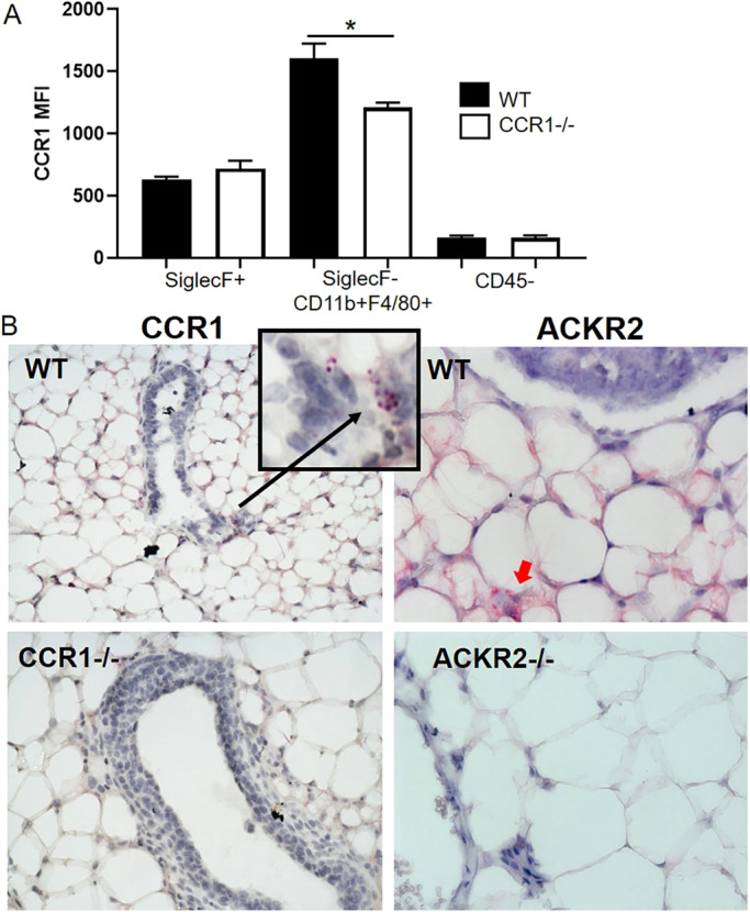
Fig. 3.

CCR1 and ACKR2 are expressed surrounding epithelium in the mammary gland. (A) Flow cytometry analysis of CCR1 expression by enzymatically digested wild-type (black bars, n=6) and Ccr1−/− (white bars, n=4) mammary gland cells: CD45+ SiglecF+, CD45+SiglecF-CD11b+F480+ and CD45−. (B) RNAscope in situ hybridisation of CCR1 (highlighted by a black arrow) and ACKR2 (highlighted by a red arrow) in the developing virgin mammary gland of WT, Ccr1−/− and Ackr2−/− mice. Significantly different results are indicated: two-tailed t-test, *P=0.0305. Data are mean±s.e.m.
