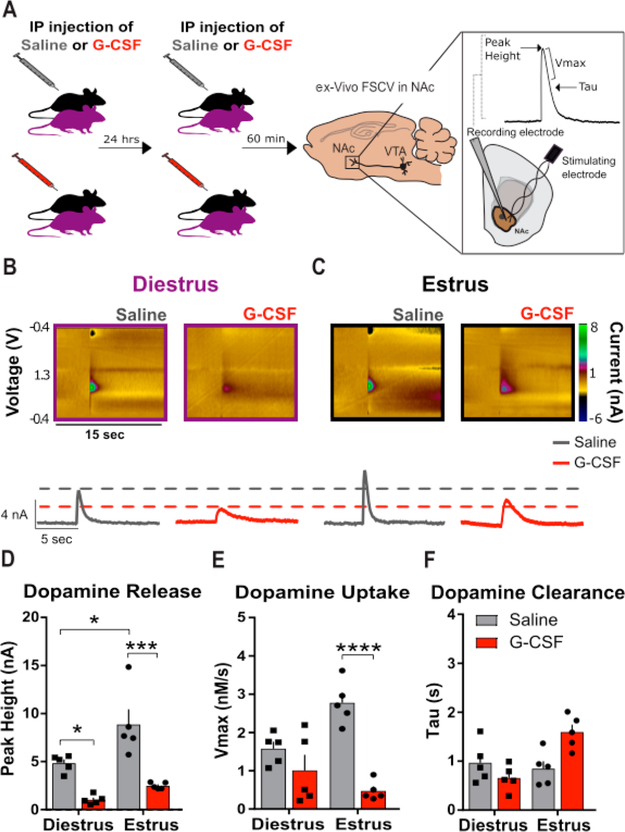
Figure 3.
G-CSF decreases presynaptic dopamine release in the nucleus accumbens of female mice. (A) Timeline of G-CSF injections. Animals were injected with either saline or G-CSF 24 h and then 60 min before ex vivo voltammetry (left; n = 5 per group) to ensure that G-CSF blood levels were elevated for a comparable time as compared to the CPP experiments. FSCV was used to record dopamine release and uptake in the NAc (right). (Inset) Peak height, Vmax, and tau measurements were used to assess the signal. (B,C) Color plots (top) and current versus time plots (bottom) showing the presence of dopamine after electrical stimulation in diestrus (B) and estrus (C) animals. (D) Group data showing enhanced dopamine release in the saline treated animals in estrus compared to diestrus, and decreased dopamine release in the G-CSF treated animals in diestrus and estrus. (E) G-CSF treatment decreased maximal rates of dopamine uptake (Vmax) only in estrus. (F) There was no difference in dopamine clearance as measured by tau. *p < 0.05, **p < 0.01, ***p < 0.0001. Data presented as mean ± SEM.

