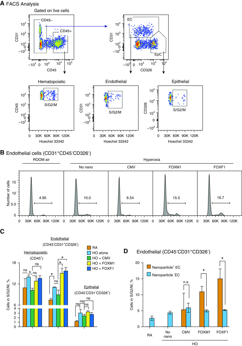
Figure 7.
Nanoparticle delivery of FOXM1 (forkhead box M1) or FOXF1 (forkhead box F1) increases endothelial cell proliferation during the recovery period after neonatal hyperoxia (HO). (A) Fluorescence-activated cell sorter (FACS) gating strategy to identify hematopoietic (CD45+CD31−), epithelial (EpC; CD326+CD45−CD31−), and endothelial cells (EC; CD31+CD45−CD326−) in mouse lung tissue. Mice were exposed to HO or room air (RA) from Postnatal Day 1 (P1) to P7, followed by RA exposure. FACS analysis of enzymatically digested lung tissue was performed 2 days after injury at P9. Dot plots show FACS analysis of cells obtained from HO-treated lungs. Hoechst 33342 dye was used to identify cells undergoing S, G2, and M phases of the cell cycle. (B and C) Histograms in B show the percentage of EC in S, G2, and M phases of the cell cycle after nanoparticle delivery of FOXM1 or FOXF1 compared with cytomegalovirus (CMV)-empty control. Data were quantitated in C and compared between different pulmonary cell types (n = 3–4 mice per group). Nanoparticle delivery of FOXM1 or FOXF1 increases the percentage of proliferating EC in HO-treated lungs. (D) Comparison of EC with and without nanoparticles. Cell proliferation is higher in EC containing nanoparticles with FOXM1 or FOXF1 (Nanoparticle+ EC) than in EC without nanoparticles (Nanoparticle− EC) (n = 3–4 mice per group). Error bars are mean ± SE. *P < 0.05. n.s. = not significant.

