Fig. 6.

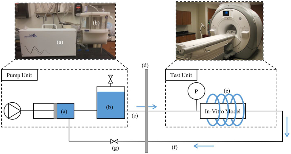
MRI experiment setup. (a) Pulsatile pump; (b) 1.5 L compliance chamber; (c) 25 ft of PVC tubing; (d) wall separating scan room and control room; (e) cardiac coil; (f) 25 ft of PVC tubing; (g) resistance valve

MRI experiment setup. (a) Pulsatile pump; (b) 1.5 L compliance chamber; (c) 25 ft of PVC tubing; (d) wall separating scan room and control room; (e) cardiac coil; (f) 25 ft of PVC tubing; (g) resistance valve