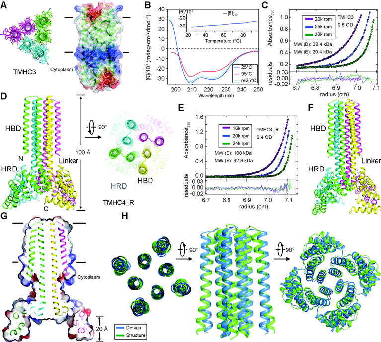
Fig. 4.
Stability and structural characterization of designs with six and eight membrane spanning helices. (A) Model of designed transmembrane trimer TMHC3 with six transmembrane helices. Stick representation from periplasmic side (left) and lateral surface view (right) are shown. (B) Circular dichroism characterization of TMHC3; the design is stable up to 95°C. (C) Representative analytical ultracentrifugation sedimentation-equilibrium curves at three different rotor speeds for TMHC3. The data fit to a single ideal species in solution with molecular weight close to that of the designed trimer. (D) Model of designed transmembrane tetramer TMHC4_R with eight transmembrane helices. The four protomers are colored green, yellow, magenta and cyan, respectively. (E) Analytical ultracentrifugation sedimentation-equilibrium curves at three different rotor speeds for TMHC4_R fit well to a single species with a measured molecular weight of ~94 kDa. (F) Crystal structure of TMHC4_R. The overall tetramer structures are very similar to the design model, with a helical bundle body and helical repeat fins. The outer helices of the transmembrane hairpins tilt off the axis by ~10°. (G) Cross section through the TMHC4_R crystal structure and electrostatic surface; the HRD forms a bowl at the base of the overall structure with a depth of ~20 Å. The transmembrane region is indicated in lines. (H) Three views of the backbone superposition of TMHC4_R crystal structure and design model.

