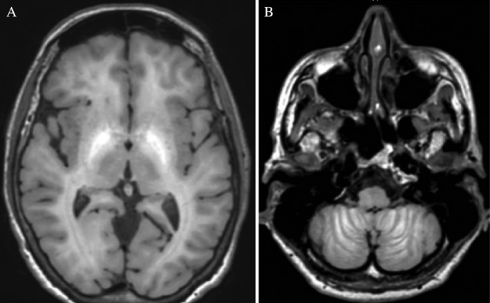
Figure 1.

Brain magnetic resonance imaging demonstrates hyperintense T1 signal of the pallidum (A) as well as in the cerebellar folds (B). To a lesser degree, hyperintense T1 signals were also observed in the subcortical white matter, anterior midbrain, and vermis. Fluid attenuated inversion recovery and susceptibility weighted images were normal.
