
Figure 6.
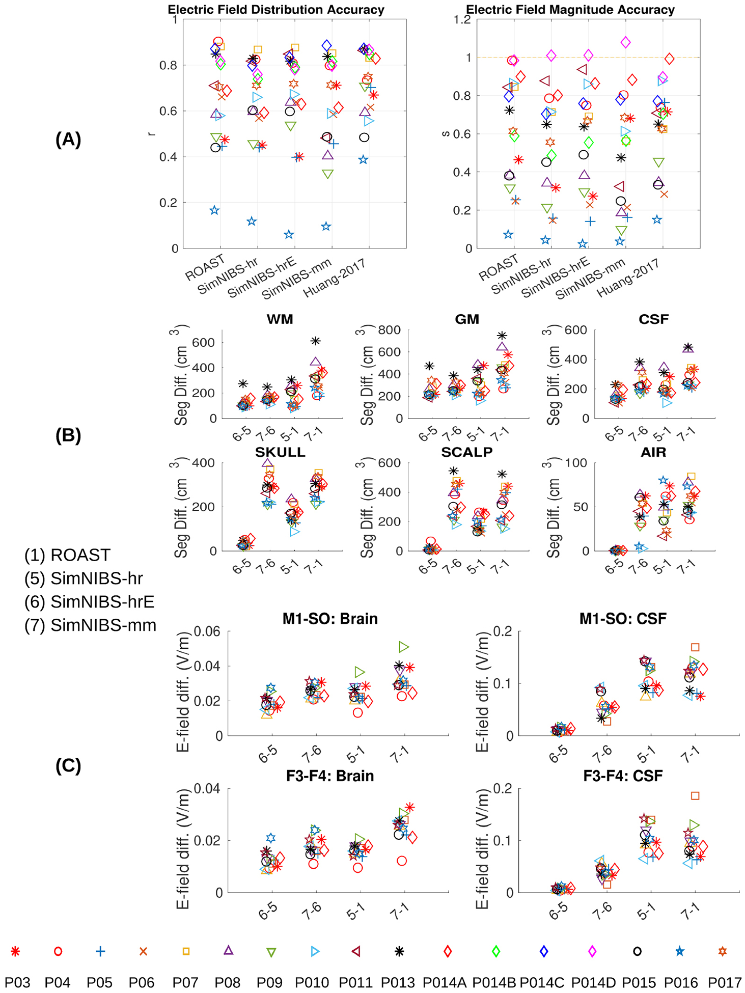
(A) Comparing the four modeling pipelines and an old published manual approach (Huang et al 2017) using intracranial electrical recordings from human subjects under transcranial stimulation as the ground truth. Left panel in (A): correlation indicates the accuracy of the spatial distribution. Right panel in (A): scaling needed to best match the estimated fields with the measured fields. Correct magnitude prediction corresponds to a scale s = 1. Also the differences between the four pipelines in terms of segmentaion and electric field distributions are shown in (B) and (C).