Figure 4.
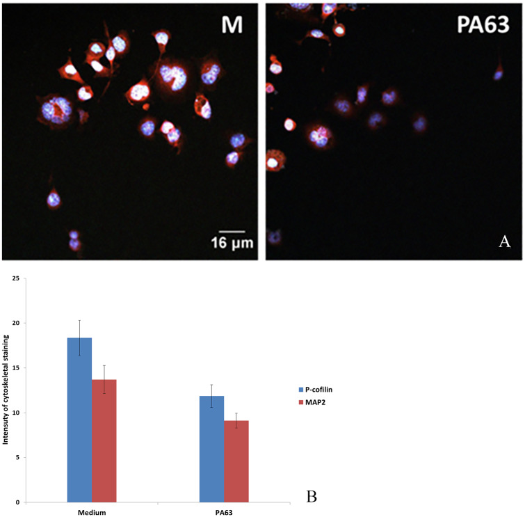
(A) N2A cells cultured in medium (M) and with PA63. Intensity of staining of MAP2 and p-cofilin combined. Cell nuclei are stained with DAPI (blue). (B) Intensity of staining for MAP2 and p-cofilin was ranked arbitrarily from 0 to 25. Intensity of staining for combined microtubular and actin networks was approximately 30% less in the presence of PA63 (B) (*P < .05). DAPI indicates 4′,6-diamidino-2-phenylindole; MAP2, microtubule-associated protein-2; N2A cells, neuroblastoma 2A cells; PA63, anthrax protective antigen 63.

