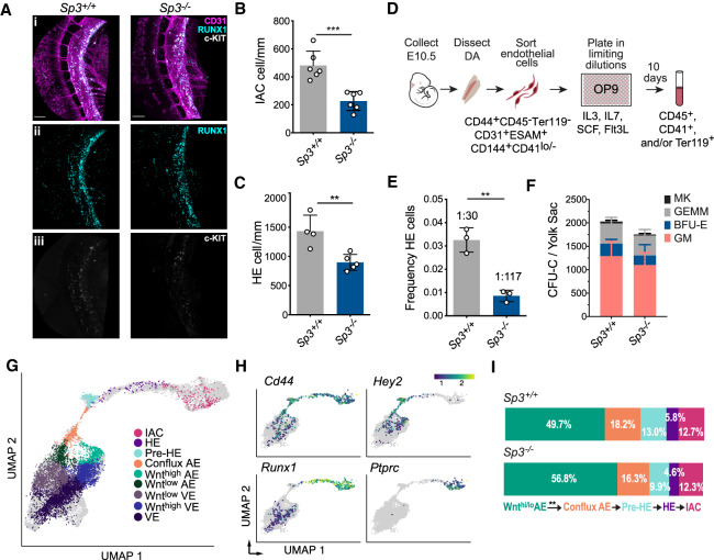
Figure 7.
SP3 regulates the frequency of HE cells in the dorsal aorta of mouse embryos. (A) Confocal z-projections (z-intervals = 2.5 µm) of dorsal aortas from E10.5 Sp3+/+ and Sp3−/− mouse embryos. Embryos were immunostained for CD31 (panel i), RUNX1 (panels i,ii) and c-KIT (panels i,iii). Scale bars, 100 µm. (B) Quantification of CD31+RUNX1+c-KIT+ intra-aortic hematopoietic cluster cells in the lumen of the dorsal aortas of E10.5 embryos (mean ± SD, unpaired two-tailed t-test). (C) Quantification of HE cells (CD31+RUNX1+c-KIT−) in the wall of the dorsal aorta (mean ± SD, unpaired two-tailed t-test). (D) Illustration of a limiting dilution assay to determine the frequency of functional HE cells in a purified population of CD44+ endothelial cells enriched for HE. (E) Frequency of HE cells in CD44+CD45−Ter119−CD31+ESAM+CD144+CD41lo/− cells purified from the dorsal aortas of E10.5 Sp3+/+ or Sp3−/− embryos (mean ± SD, unpaired two-tailed t-test). Each replicate consisted of pooled cells from separate litters of E10.5 embryos collected in independent experiments. (***) P ≤ 0.001; (**) P ≤ 0.01. (F) Colony-forming unit cells (CFU-Cs) per E10.5 YS (mean ± SD). (MK) Megakaryocyte; (GEMM) granulocyte macrophage–erythroid–megakaryocyte; (BFU-E) erythroid blast-forming unit; (GM) granulocyte macrophage. Data are from three independent litters. (G) Projection of combined Sp3+/+ and Sp3−/− cells onto the EHT trajectory from Zhu et al. (2020). Cell types were annotated using a k-nearest-neighbor classifier (see Supplemental Material). AE is defined by gene module scoring of arterial-specific genes (Efnb2, Nrp1, Gja5, Bmx, Notch1, Notch4, Dll4, Jag1, Jag2, Acvrl1, Epas1, 8430408G22Rik, and Vegfc) relative to venous-specific genes (Ephb4, Flt4, Nrp2, Nr2f2, and Emcn) as described in Zhu et al. (2020). (H) Expression pattern of selected cell type-specific genes on the UMAP plot. (I) Fraction of Wnthigh/low AE, conflux AE, pre-HE, HE, and IAC cells in Ter119−CD41lo/−CD31+CD144+ESAM+ cells purified from E10.5 Sp3+/+ versus Sp3−/− embryos. Only the difference in the proportion of cells in Wnthigh/low AE versus conflux AE was significant (P < 0.006, proportion test).

