
Figure 1.
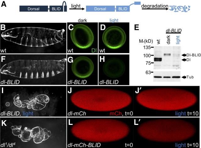
Illumination with blue light induces degradation of Dl-BLID fusion proteins. (A) The Dl-BLID construct. Blue-light illumination causes a degradation sequence to be exposed, resulting in the degradation of the entire fusion protein. (B,F,I,K) Cuticle preparations of embryos derived from wild-type mothers without illumination (B; n = 181/190), dl-BLID mothers without illumination (F; n = 147/310), dl-BLID mothers with 4 h of blue LED illumination (I; n = 31/36), and dl null mutant (dl1/dl4) mothers without illumination (K; 142/142). (C,D,G,H) Manually crossed sectioned embryos stained with anti-Dl antibody (green) derived from wild-type mothers without illumination (C; n = 5/5), wild-type mothers with 1 h of blue LED illumination (D; n = 5/5), dl-BLID mothers without illumination (G; n = 5/5), and dl-BLID mothers with 1 h of blue LED illumination (H; n = 4/5). All embryos in C,D,G,H were imaged at the same settings, demonstrating a clear decrease in Dl levels in H. (E) Western blot of wild type (lane 1), dl-BLID without illumination (lane 2), and dl-BLID with 30-min blue LED illumination (lane 3). Top blot is probed with anti-Dl antibody. Bottom blot is probed with anti-Tubulin antibody to serve as a loading control. Arrows indicate the approximate locations of Dl, Dl-BLID, and Tubulin bands. (J,J′,L,L′) Snapshots from live imaging movies of dl-mCherry (n = 1) and dl-mCherry-BLID (n = 3) at the start (J,L; t = 0) and after 10 min of 40% power blue laser illumination (J′,L′; t = 10). All embryos/larval cuticles are oriented with anterior to left and dorsal up, except cross-sections, which are oriented with the ventral side at the bottom and the dorsal side at the top.