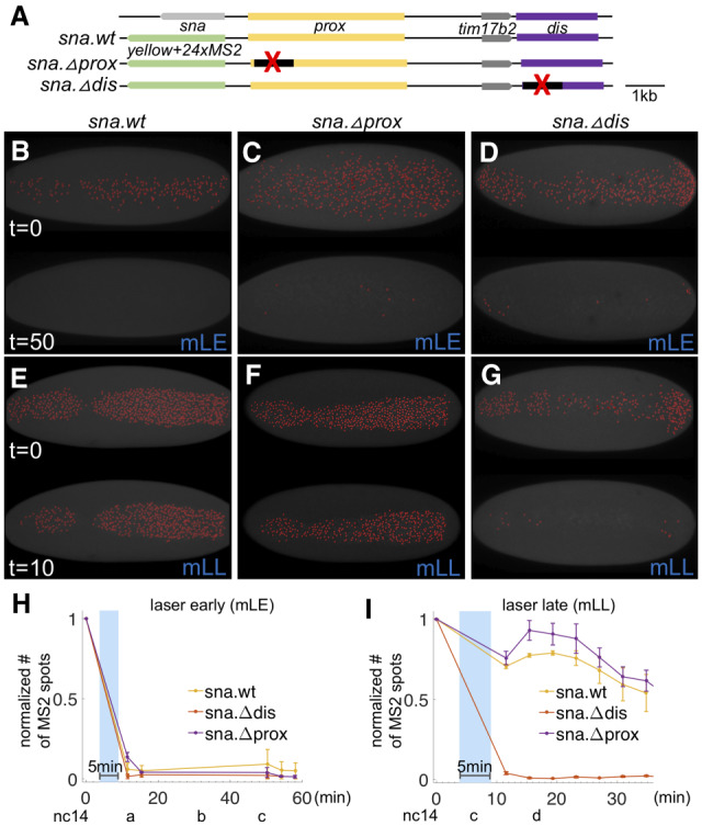
Figure 3.

High levels of Dl are required for sna activativation only at early stages, but not at late stages, in which sna expression is predominantly supported by the sna distal enhancer. (A) Scheme of large reporter constructs used to assay sna transcriptional activities by MS2-MCP system (Bothma et al. 2015). (B–G) MCP.GFP signals associated with the sna MS2 reporter were imaged (false-colored red dots) in dl-BLID with early (B–D) or late (E–G) blue laser illumination that is MS2-MCP imaging compatible (“mLE” and “mLL,” respectively) (see also Supplemental Fig. S3) in various sna regulatory conditions including wildtype (sna.wt; B,E), proximal enhancer deletion (sna.Δprox; C,F), and distal enhancer deletion (sna.Δdis; D,G). Images are snapshots from movies, before illumination (top) and after illumination (bottom) of each panel. Three movies were taken for each condition. Ventral views of embryos are shown with anterior oriented to the left. (H,I) Quantitative analysis of the number of MCP.GFP dots associated with the sna MS2 reporter in dl-BLID embryos with sna.wt, sna.Δprox, or sna.Δdis sna regulatory condition. Number of MS2-MCP.GFP spots are counted in each time frame, and the values are normalized to the initial value detected in the first frame (before 5 min blue-laser illumination with 15% laser power) with early laser (H) or late laser (I) illumination. Blue shade indicates a time frame of 5-min 15% blue-laser illumination. Error bars represent standard error of the mean. For individual traces, see Supplemental Figure S5. For details for detection of sna.MS2-MCP.GFP and blue-laser illumination, see Supplemental Figure S3.
