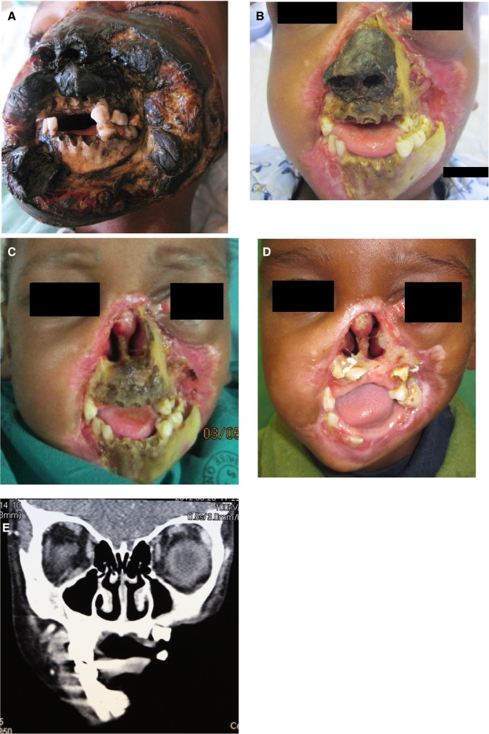
Figure 7.

(A) Severe necrotic destruction of the nose, lips, and cheeks extending to the infra‐orbital margin, and denudation and necrosis of the anterior mandible and anterior maxilla of a 6‐y‐old HIV‐seropositive highly active antiretroviral treatment‐naïve child with a CD4 + T cell count of 6 cells/mm3. (B) The appearance 2 wk after hospital admission: much of the necrotic soft tissue has been removed. (C) All the necrotic soft tissue removed. (D) The appearance 3 mo after resection of all the necrotic soft tissue and bone with extensive scarring around the defect. (E) Computed tomography image showing the extent of the damage to the mandible and maxilla (Head Neck Pathol 2013; 7:188‐192, reproduced with permission8)
