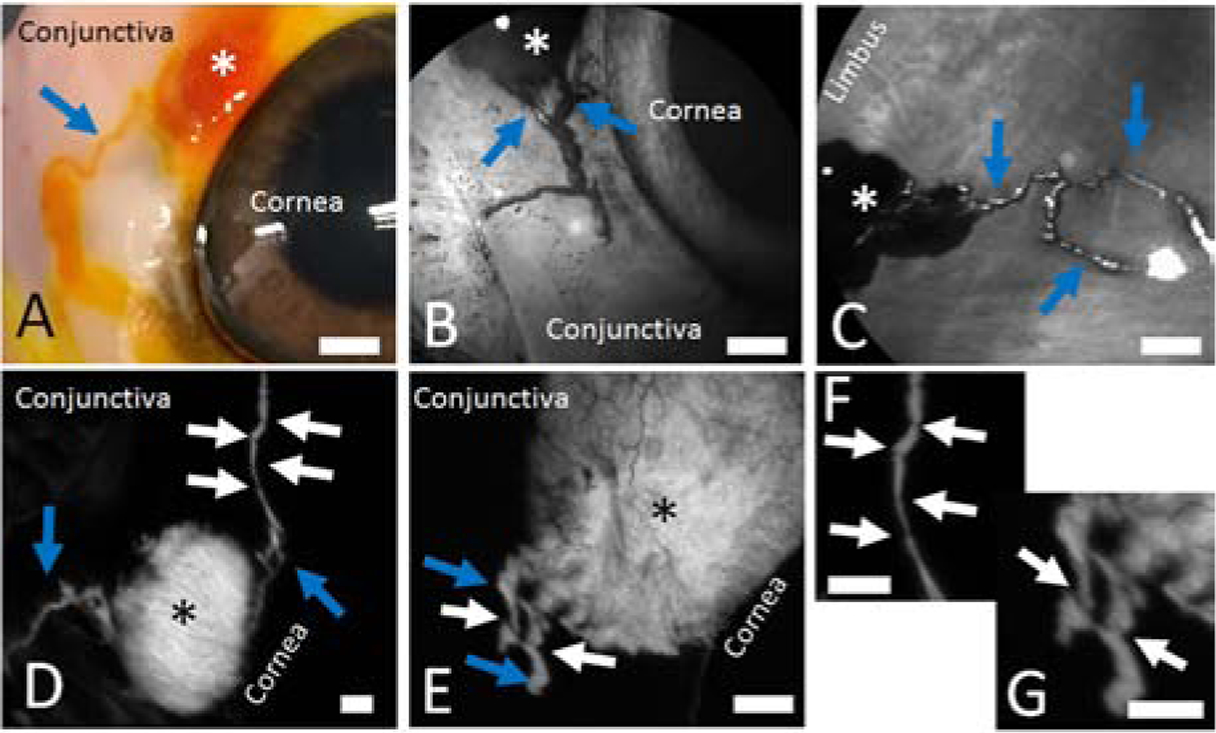
Figure 1.

Distinct Outflow Pathways Can Be Visualized off Blebs Created Using Multiple Tracers in Multiple Species.
In all cases, 100 microliters of a tracer are injected subconjunctival using a 30-gauge needle to create a bleb (asterisks). Distinct outflow pathways are seen arising off of the blebs. A/B/D) Porcine eyes. C) Bovine eye. E) Enucleated right eye from a 54-year-old male. Blue arrows demonstrate outflow pathways. White arrows show intervening regions in the outflow pathways with narrow bands of darker signal. F/G) Zoomed in images of D and E, respectively, showing the dark bands more clearly (white arrows). Scale bars = 1 mm.
