
Figure 5.
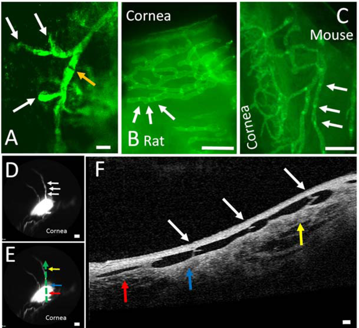
Structural Characteristics of Outflow Pathways Off Blebs Are Reminiscent of Lymphatics.
A) Two-photon microscopy image of fluorescent dextrans trapped in porcine outflow pathways showed structures that are similar to blind-end lymphatics capillaries (white arrow) leading to a possible initial lymphatic trunk with signal variation along the way which may represent valves (orange arrow) (scale bar = 200 microns). This region came from the same tissue as Figure 1D and Figure 3B that included the outflow pathway but not the bleb. Natively fluorescent conjunctival lymphatics are seen from transgenic (B) rat and (C) mouse that expresses EGFP under a Prox-1 promoter. B/C) These lymphatics showed bright spots (white arrows) that are known to represent lymphatic valves (scale bars = 1 mm). D) In a porcine eye, a bleb was created with two outflow pathways arising superior (scale bar = 1 mm). E) OCT was performed and placed on the outflow pathway as denoted by the dotted green arrow with the direction of flow from red to blue to yellow horizontal arrows (scale bar = 1 mm). F) Bicuspid valves (white arrows; that match white arrows in D) are seen in the direction of flow (red to blue to yellow vertical arrows) (scale bar = 100 microns). The red/blue/yellow arrows in (E) match the same colored arrows in (F) and demonstrate a 2-dimensional representation of the outflow pathway off of the bleb.