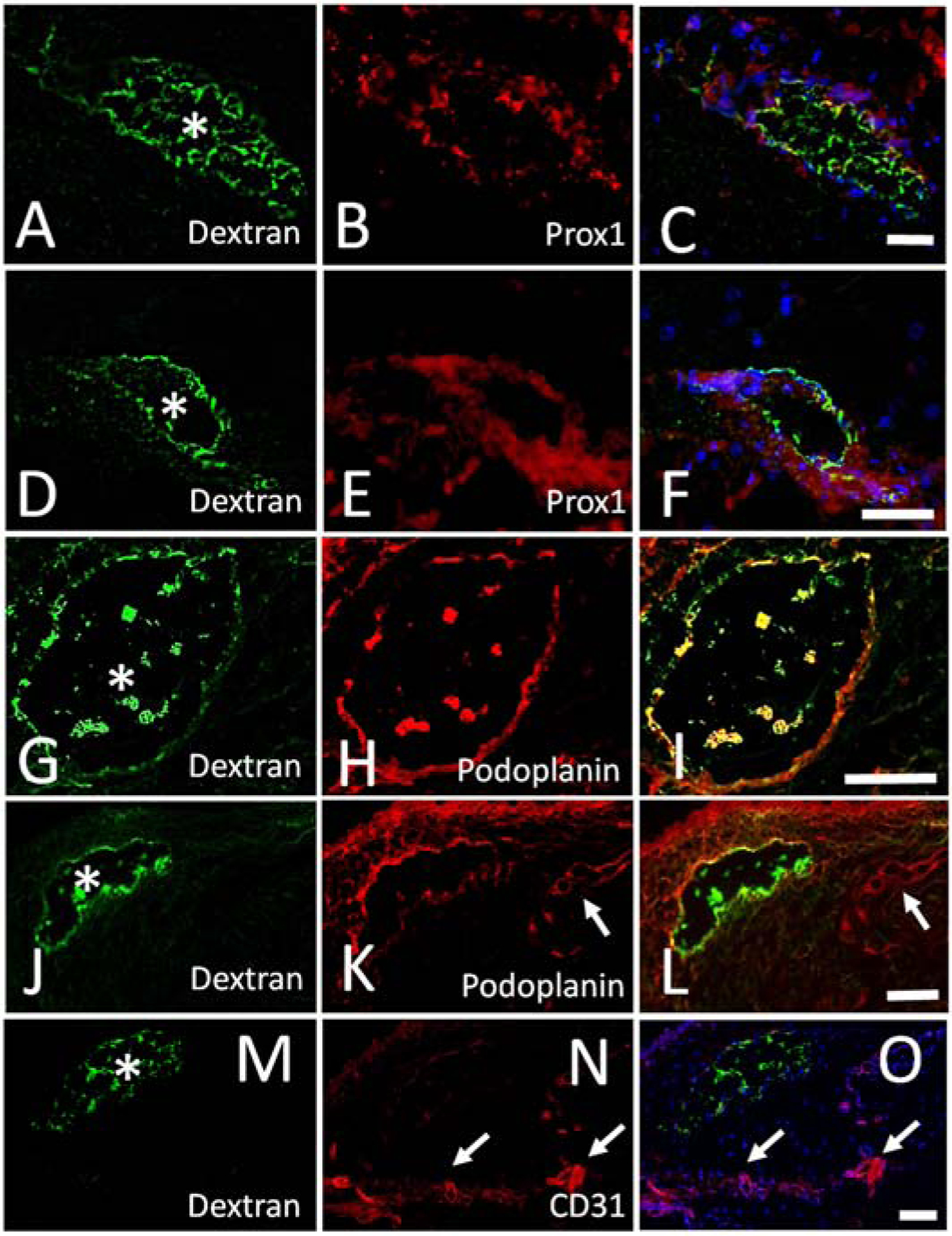
Figure 6.

Outflow Pathways Off Blebs Express Lymphatic Markers in Porcine Eyes.
A/D/G/J/M) After injection of a fluorescent dextran bleb, outflow pathways were seen, fixed, and sectioned. Trapped tracer was seen lining the outflow pathway lumens (white asterisks). A/D) Immunolabeling on the same section against (B/E) Prox-1 showed (C/F) co-localization with the tracer-labeled outflow pathways. G/J) Immunolabeling on the same section against (H/K) podoplanin showed (I/L) co-localization with the tracer-labeled outflow pathways. K/L) Shows images with dextran and podoplanin co-localization but also another lymphatic (white arrow) that did not show tracer labeling. M-O) Immunolabeling on the same section against a blood vessel marker (CD31) did not show co-localization with the tracer-labeled outflow pathway but instead showed distinct blood vessels elsewhere (white arrows). Scale bars = 100 microns.
