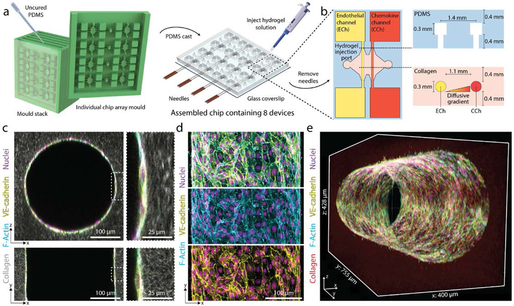
Figure 1 |. Multiplexed angiogenesis-on-a-chip platform.

a, PDMS replica casts from 3D-printed moulds are bonded to glass coverslips. Each chip is composed of a 2×4 array of single devices. A pair of needles are inserted into each device, and type I collagen solution is injected into each device and gelled around needles. Hollow channels are generated upon needle removal. b, Inserted needles are suspended above the glass coverslip bottom and below the PDMS housing to form 3D channels fully embedded within a collagen hydrogel. Each device is composed of two parallel channels. The endothelial channel (ECh) is seeded with endothelial cells to form the parent vessel. Pro-angiogenic factors are added to the chemokine channel (CCh) and form a diffusive gradient to promote 3D endothelial cell invasion across the collagen hydrogel. c, Representative images of x-z (top) and x-y (bottom) orthogonal views of parent vessels formed within fluorescently labeled 3 mg ml−1 collagen hydrogel 24-hours post-seeding. Insets indicated with dashed white lines. d, Representative images of x-y (max intensity projection) formed within a 3 mg ml−1 collagen hydrogel 24-hours post-seeding. Merge (top), F-actin (middle), VE-cadherin (bottom). e, 3D rendering of parent vessel formed within fluorescently labeled 3mg ml−1 collagen hydrogel.
