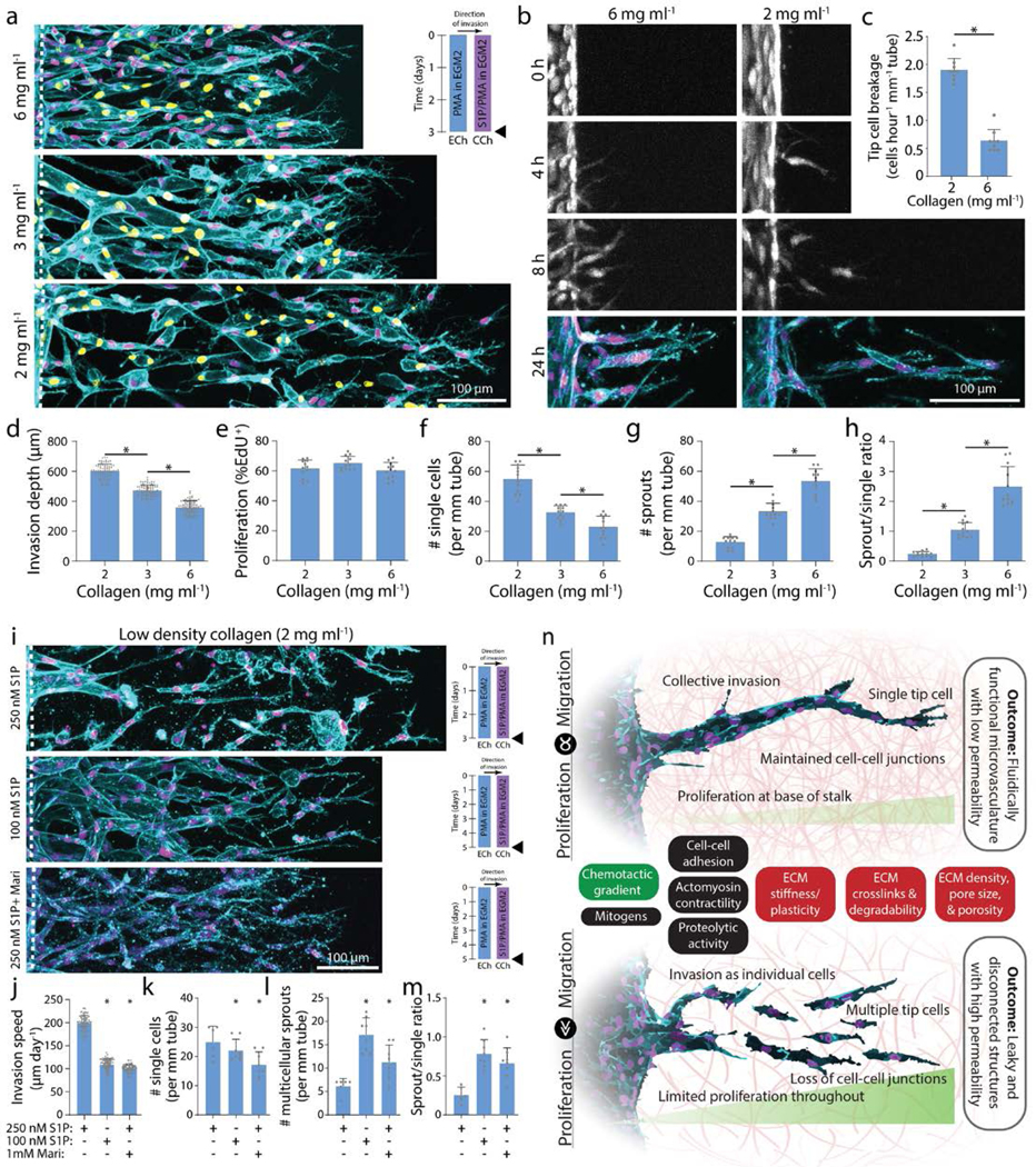
Figure 6 |. Increasing collagen density decreases invasion speed and enhances multicellular sprouting.

a, Representative images (max intensity projection) of invading endothelial cells in response to varying collagen density with S250:P25. UEA (cyan), nucleus (magenta), EdU (yellow). b, Representative time course images (max intensity projection) of invading endothelial cells (labeled with cell tracker dye) in response to varying collagen density with S250:P25. F-actin (cyan) and nucleus (magenta). c, Quantification of the frequency of tip cell breakage events from the parent vessel. d-h, Quantifications of invasion depth, proliferation, and morphology of invading endothelial cells as single cells or multicellular sprouts from conditions in (a). i, Representative images (max intensity projection) of invading endothelial cells in 2 mg ml−1 collagen with control (S250:P25), decreased S1P (S100:P25), and MMP inhibition (S250:P25). F-actin (cyan) and nucleus (magenta). j-m, Quantifications of invasion speed, and morphology of invading endothelial cells as single cells or multicellular sprouts. All data presented as mean ± s.d.; * indicates a statically significant comparison with P<0.05 (two-tailed Student’s t-test (c, j-m) and one-way analysis of variance (d-h)). For tip cell breakage analysis (c), n=9 vessel segments (each 400 μm length) per condition. For invasion depth analysis (d), n≥80 vessel segments (each 100 μm length) per condition. For proliferation and migration mode analysis (e-h, k-m), n≥6 vessel segments (each 800 μm length) per condition from n=2 devices/condition (technical replicates) over n≥3 independent studies (biological replicates). For invasion speed analysis (j), n≥81 vessel segments (each 100 μm length) per condition. n, Schematic illustration highlighting the relationship between endothelial cell migration and proliferation on invasion morphology as multicellular sprouts (top; balanced migration and proliferation) vs single cells (bottom; excessive migration or insufficient proliferation). Soluble (green) and physical extracellular matrix (red) cues influence this balance in addition to other potential cell functions (black) that may regulate multicellular sprouting and formation of functional microvasculature.
