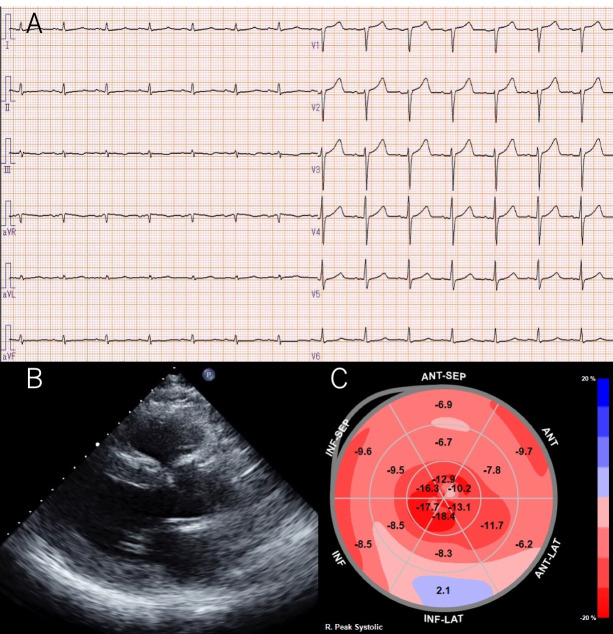
Figure 1.
(A) A 12-lead ECG shows sinus rhythm with a heart rate of 73 beats/min and decreased voltage in the limb leads. (B) Transthoracic echocardiography reveals increased left ventricular wall thickness (intraventricular septum, 16 mm; posterior wall thickness, 16 mm). (C) Two-dimensional speckle-tracking echocardiography reveals a relative apical longitudinal strain (LS) (=average apical LS/average basal LS+mid LS) of 1.9 with apical sparing.

